
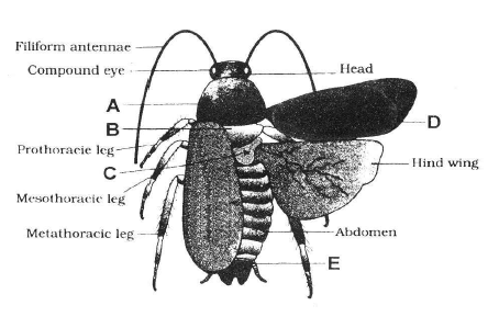
The following figure shows the extarnal features of cockroach with few structures labelled as A, B, C, D, & E. Identify A to E .
A- Mesothorax, B-Pronotum, C-Metathorax, D-Tegmina, E-Anal style
A- Pronotum, B-Metathorax, C-Mesothorax, D-Tegmina, E-Sterna
A- Pronotum, B-Mesothorax, C-Metathorax, D-Tegmina, E-Anal cerci
A- Pronotum, B-Mesothorax, C-Metathorax, D-Tegmina, E-Anal style
Correct Answer :
C. A- Pronotum, B-Mesothorax, C-Metathorax, D-Tegmina, E-Anal cerci
A - Pronotum; B - Mesothorax; C - Metathorax; D - Tegmina; E - Anal cerci
Related Questions
Four healthy people in their twenties got involved in injuries resulting in damage and death of a few cells of the following.
Which of the cells are least likely to be replaced by new cells?
Liver cells
Neurons
Malpighian layer of the skin
Osteocytes
The only type of cell seen in a tendon is
muscle fibres
reticular cells
collagenous cells
fibroblasts
Statement 1 : Cartilage (protein matrix) and bone (calcium matrix) are rigid connective tissue.
Statement 2 : Blood is connective tissue in which plasma is the matrix.
Statement- 1 and statement-2 are true and statement-2 is a correct explanation for statement -1
Statement -1 and statement-2 are true and statement-2 is not a correct explanation for statement -1
Statement - 1 is true and statement- 2 is false
Both the statements are false.
What external changes are visible after the last moult of a cockroach nymph?
Development of anal cerci.
Development of both forewings and hind wings.
Development labium.
Mandibles become harder.
The mouth parts of cockroach are
cutting and biting type.
piercing and sucking type.
sucking and rasping type.
sucking and siphoning type.
In all connective tissues, except which of the following, the cells secretes the fibres of collagen or elastin protein ?
Bone
Cartilage
Areolar connective tissue
Fluid connective tissue
Match column-I with column-II and choose the correct option.
| Column-I | Column-II |
|---|---|
| A. Periplaneta | I. Hepatic caecae americana |
| B. A ring of 6-8 blind | II. Phylum arthropoda tubules |
| C. Vascular system | III. Spiracles |
| D. 10 pairs of small | IV. Malpighian tubules holes |
| E. Excretion | V. Open type |
A I; B II; C III; D IV; E V
A II; B I; C V; D III; E IV
A II; B I; C III; D V; E IV
A III; B IV; C II; D V; E I
The fibres of which of the following muscles are fusiform and do not show striations
Skeletal muscles
Cardiac muscles
Both (a) and (b)
Smooth muscles
Match the types of connective tissue given in column-I with their examples given column-II and choose the correct option.
| Column-I | Column-II |
|---|---|
| (Types of connective) | (Examples) |
| A. Loose connective | I. Tendons and ligaments tissue |
| B. Dense regular | II. Skin tissue |
| C. Dense irregular | III. Cartilage, bones, blood tissue |
| D. Specialized | IV. Fibroblasts, macrophages connective tissue and mast cells |
A I; B IV; C II; D III
A I; B IV; C III; D II
A IV; B I; C II; D III
A IV; B II; C I; D III
The blood of cockroach contains no respiratory pigment. It means that
cockroach does not respire.
respiration is anaerobic.
oxygen goes directly into tissues by diffusion.
oxygen goes directly into tissues by intracellular capillary system.
Hair present in the skin are
epidermal in origin and made of dead cells.
epidermal in origin and made of living cells.
dermal in origin and made of living cells.
dermal in origin and made of dead cells.
Figure given below shows reproductive system of female cockroach. The correct labellings indicated by alphabets (A, B & C) are respectively
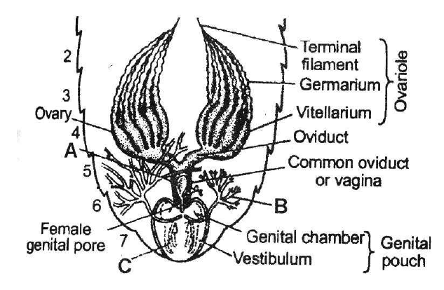
A-Spermatheca, B-Collaterial glands, C-Gonapophyses
A-Phallic gland, B-Collaterial glands, C-Gonapophyses
A-Spermatheca, B-Seminal vesicle, C-Gonapophyses
A-Spermatheca, B-Collateral glands, C-Tegmina
If the head of cockroach is cut off, it will still alive for as long as one week. It is because of
the body which is covered with a hard chitinous exoskeleton.
head which holds a bit of nervous system.
head which is of no use.
food capturing appratus which is found elsewhere.
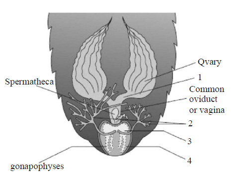
Refer the given figure of female reproductive system of cockroach and identify the correct labels (marked as 1, 2, 3 and 4) which are collectively called genital pouch.
1 & 2
1 & 3
2 & 4
3 & 4
Which of the follwing statement(s) is/are correct ?
- Cockroaches are brown or black bodied animals that are included in class insecta of phylum arthropoda.
- Males bear a pair of short, thread like anal styles which are absent in females.
- Heart of cockroach consists of elongated muscular tube lying along mid dorsal line of thorax and abdomen.
- The nymph grows by moulting about 13 times to reach the adult form.
Only (i)
Both (ii) and (iii)
Both (i) and (ii)
All of the above
When cardiac muscle cells are damaged by a heart attack, they are usually replaced by
connective tissue cells
new smooth muscle cells
new cardiac muscle cells
epithelial cells
Statement 1: Typhlosole increases the effective area of absorption in the intestine.
Statement 2: Typhlosole, present in the intestine, is the characteristic feature of cockroach.
Statement 1 and 2 are true and statement 2 is the correct explanation of statement 1.
Statement 1 and 2 are true and statement 2 is not the correct explanation of statement 1.
Both statements 1 and 2 are false.
Statement 1 is true and statement 2 is false.
Which of the following statement(s) is/are correct regarding cockroaches ?
- The body of the cockroach is segmented and divisible into three distinct regions head, thorax and abdomen.
- Blood vascular system is of closed type.
- They are monoecious and both sexes have well developed reproductive organs.
- The development of P. americana is paurometabolus, meaning there is development through nymphal stage.
Both (i) and (ii)
Both (ii) and (iii)
Both (i) and (iv)
All of these
Which of the following type of cell junction is not found in animal tissues ?
Adhering junction
Tight junction
Gap junction
Plasmodesmata
Epithelial tissue is distinguished from connective tissue, muscular, or nervous tissue by its
large extracellular matrix.
contractibility.
ability to carry action potentials.
basement membrane.
The ciliated columnar epithelial cells in humans occur in
Eustachian tube and stomach lining
bronchioles and fallopian tubes
bile duct and oesophagus
fallopian tubes and urethra
The following figure shows the extarnal features of cockroach with few structures labelled as A, B, C, D, & E. Identify A to E .
A- Mesothorax, B-Pronotum, C-Metathorax, D-Tegmina, E-Anal style
A- Pronotum, B-Metathorax, C-Mesothorax, D-Tegmina, E-Sterna
A- Pronotum, B-Mesothorax, C-Metathorax, D-Tegmina, E-Anal cerci
A- Pronotum, B-Mesothorax, C-Metathorax, D-Tegmina, E-Anal style
Match the epithetial tissue given in column-I with its location given in column-II and choose the correct option.
| Column I | Column II |
|---|---|
| (Epithelial tissue) | (Location) |
| A. Cuboidal | I. Epidermis of skin |
| B. Ciliated | II. Inner lining of blood vessels |
| C. Columnar | III. Inner surface of gall bladder |
| D. Squamous | IV. Inner lining of fallopian tube |
| E. Keratinized | V. Lining of pancreatic duct squamous |
A V; B IV; C II; D III; E I
A III; B IV; C V; D II; E I
A V; B IV; C III; D II; E I
A III; B IV; C V; D I; E II
Select the correct statement regarding Periplaneta americana
There are 16 very long malpighian tubules present at the junctions of midgut and hindgut.
Grinding of food is carried out only by the mouth parts.
Nervous system is located ventrally and consists of segmentally arranged ganglia joined by a pair of longitudinal connectives.
Females bear a pair of short thread like anal styles.
The intercellular material of the given figure is solid and resists compression. Identify the figure and the label marked as A & B.
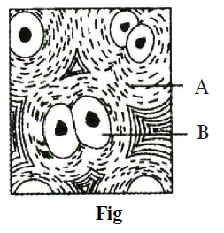
Fig - Cartilage, A - Collagen, B - Collagen
Fig - Cartilage, A - Microtubule, B - Collagen fibres
Fig - Bone, A - Chondrocyte, B - Chondroclast
Fig - Bone, A - Chondroclast, B - Osteoblast
The given figure shows the digestive system of cockroach with few structures marked as A, B, C and D. Identify structures A to D.
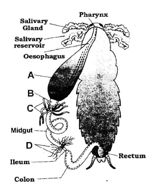
A-Gizzard, B-Crop, C-Hepatic caecae, D-Malpighian tubules
A-Crop, B-Gizzard, C-Hepatic caecae, D-Malpighian tubules
A- Crop, B-Gizzard, C-Malpighian tubules, D-Hepatic caecae
A- Gizzard, B-Crop, C-Malpighian tubules, D-Hepatic caecae
In cockroach head can move in all directions due to
absence of neck.
fusion of all 6 segments of head.
flexible neck.
head is small and light weight.
Three essential components of most neurons are
simple epithelium, extracellular matrix and nerves.
axon, dendrites and cell body.
nerve cells, synapse and neuroglia.
mylein sheeth, node of Ranvier and Schwann cells.
The following figures A, B and C are types of muscle tissue. Identify A, B and C.
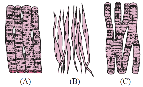
A Smooth muscle, B Cardiac muscle, C Skeletal muscle
A Skeletal muscle, B Smooth muscle, C Cardiac muscle
A Cardiac muscle, B Smooth muscle, C Skeletal muscle
A Smooth muscle, B Skeletal muscle , C Cardiac muscle
Which of the following statement(s) is/are correct about muscle tissue ?
Each muscle is made of many long, cylindrical fibres arranged in parallel arrays.
Muscle fibres contract (shorten) in response to stimulation, then relax (lengthen) and return to their uncontracted state in a coordinated fashion.
Muscles play an active role in all movements of the body.
All of the above