
Three essential components of most neurons are
simple epithelium, extracellular matrix and nerves.
axon, dendrites and cell body.
nerve cells, synapse and neuroglia.
mylein sheeth, node of Ranvier and Schwann cells.
Correct Answer :
B. axon, dendrites and cell body.
A neuron (also known as a neurone or nerve cell) is an electrically excitable cell that processes and transmits information through electrical and chemical signals. These signals between neurons occur via synapses, specialized connections with other cells. A typical neuron possesses a cell body (soma), dendrites, and an axon.
Related Questions
The following figure shows the extarnal features of cockroach with few structures labelled as A, B, C, D, & E. Identify A to E .
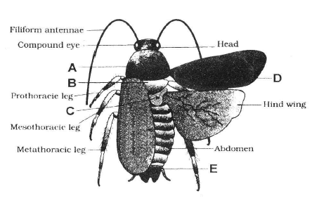
A- Mesothorax, B-Pronotum, C-Metathorax, D-Tegmina, E-Anal style
A- Pronotum, B-Metathorax, C-Mesothorax, D-Tegmina, E-Sterna
A- Pronotum, B-Mesothorax, C-Metathorax, D-Tegmina, E-Anal cerci
A- Pronotum, B-Mesothorax, C-Metathorax, D-Tegmina, E-Anal style
Three essential components of most neurons are
simple epithelium, extracellular matrix and nerves.
axon, dendrites and cell body.
nerve cells, synapse and neuroglia.
mylein sheeth, node of Ranvier and Schwann cells.
Intercalated discs are the communication junctions between the cells of
cardiac muscles
striped muscles
adipose tissue
nerve and striated muscles
Refer the given figure of female reproductive system of cockroach and identify the correct labels (marked as 1, 2, 3 and 4) which are collectively called genital pouch.
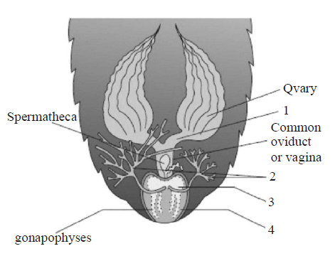
1 & 2
1 & 3
2 & 4
3 & 4
What are the three basic components of connective tissues?
Ground substance, cells and basement membrane
Cartilage, intercellular matrix and serum
Cells, protein fibers and ground substance
Collagen, elastin and reticular fibers
Which of the following type of tissue is being described by the given statements ?
- They are named because of their special function of linking and supporting other tissues/organs of the body.
- They include cartilage, bone, adipose and blood.
- They provide strength, elasticity and flexibility to the tissue.
- They also secrete modified polysaccharides, which accumulate between cells and fibres and act as matrix.
Epithelial tissue
Connective tissue
Muscle tissue
Neural tissue
Find the incorrectly matched pair.
Unicellular glandular cells - Goblet cell
Saliva - Exocrine secretion
Fusiform fibres - Smooth muscle
Cartilage - Areolar tissue
Which of the following statement(s) is/are correct regarding excretory system of cockroach ?
- Excretion is performed by malphigian tubules.
- They absorb nitrogenous waste products and convert them into uric acid which is excreted out through hindgut. Hence, this insect called ammonetelic.
- In addition, fat body, nephrocytes and uricose glands also help in excretion.
Only (i)
Both (ii) and (iii)
Both (i) and (iii)
All of these
Read the following statements.
- It is a contractile tissue present only in the heart.
- Cell junctions fuse the plasma membranes of these cells and make them stick together.
- Communication juntions at some fusion points allow the cells to contract as a unit, i.e., when one cell receives a signal to contract, its neighbours are also stimulated to contract.
Which of the following type of tissue is being described by the above statements ?
Skeletal muscle
Cardiac muscle
Smooth muscle
Cartilage
What external changes are visible after the last moult of a cockroach nymph?
Development of anal cerci.
Development of both forewings and hind wings.
Development labium.
Mandibles become harder.
In all connective tissues, except which of the following, the cells secretes the fibres of collagen or elastin protein ?
Bone
Cartilage
Areolar connective tissue
Fluid connective tissue
The secretions of endocrine glands are released directly
into the skin surface
into the blood stream
into a gland duct
into the brain tissue
If the head of cockroach is cut off, it will still alive for as long as one week. It is because of
the body which is covered with a hard chitinous exoskeleton.
head which holds a bit of nervous system.
head which is of no use.
food capturing appratus which is found elsewhere.
Nervous tissue cells that play several supporting roles but do not transmit impulses are called
glial cells
dendrites
nerve cells
neurons
Choose the correct sequence of alimentary canal of Cockroach
Gizzard -> Crop ->?Malphigian tubules ->?Hepatic caeca ->?Rectum.
Gizzard -> Hepatic caeca -> Crop -> Rectum -> Malphigian tubules.
Crop -> Gizzard ->?Hepatic caeca ->?Malphigian tubules ->?Rectum.
Crop -> Hepatic caeca ->?Gizzard -> Rectum -> Malphigian tubules.
The shape of a persons ear is due to mainly to
dense regular connective tissue
dense irregular connective tissue
elastic cartilage
fibrocartilage
Mast cells are associated with
exocrine glands
endocrine glands
areolar connective tissue
neural tissue
The mouth parts of cockroach are
cutting and biting type.
piercing and sucking type.
sucking and rasping type.
sucking and siphoning type.
Muscle tissue cells are contractile, which means they
are responsible for the production and secretion of enzymes.
are specialized in contraction and relaxation.
help in the movement of involuntary organs only.
all of the above
A student was given a sample of two tissues. He observes the tissues under the microscope and draws their figures (1 and 2) as shown below.
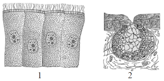
Identify the tissues (1 and 2).
1: Columnar cells bearing cilia; 2: Unicellular glandular epithelium
1: Cuboidal cells bearing cilia; 2: Multicellular glandular epithelium
1: Compound cells bearing cilia; 2: Unicellular glandular epithelium
1: Columnar cells bearing cilia; 2: Multicellular glandular epithelium
Which of the following statement(s) regarding cell junctions is/are correct ?
Tight junctions help to stop substances from leaking across a tissue.
Adhering junctions perform cementing to keep neighbouring cells together.
Gap junctions facilitate the cells to communicate with each other by connecting the cytoplasm of adjoining cells, for rapid transfer of ions, small molecules and sometimes big molecules.
All of the above
The major functions of loose connective tissue include
occupying spaces between organs and supporting epithelia.
supporting and surrounding blood vessels and nerves
cushioning organs, storing lipids and facilitating diffusion.
All of the above
Match the types of connective tissue given in column-I with their examples given column-II and choose the correct option.
| Column-I | Column-II |
|---|---|
| (Types of connective) | (Examples) |
| A. Loose connective | I. Tendons and ligaments tissue |
| B. Dense regular | II. Skin tissue |
| C. Dense irregular | III. Cartilage, bones, blood tissue |
| D. Specialized | IV. Fibroblasts, macrophages connective tissue and mast cells |
A I; B IV; C II; D III
A I; B IV; C III; D II
A IV; B I; C II; D III
A IV; B II; C I; D III
In the given diagram of areolar connective tissue, the different cells and parts have been marked by alphabets (A, B, C & D). Choose the answer in which these alphabets correctly match with the parts and cells they indicate.
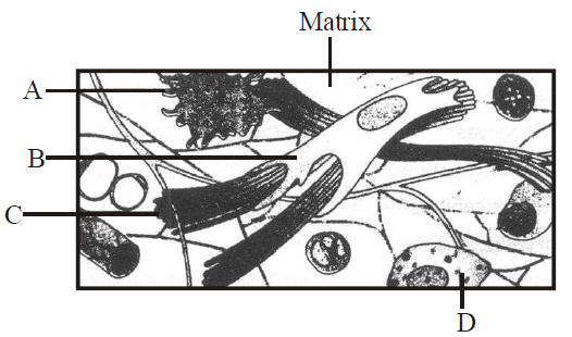
A-Adipocyte, B-Collagen fibres, C-Microfilament, D-Mast cells
A-Macrophage, B-Collagen fibres, C-Microfilament, D-Mast cells
A-Macrophage, B-Collagen fibres, C-Microtubule, D-RBC
A-Macrophage, B-Fibroblast, C-Collagen fibres, D-Mast cells
Which of the following statement(s) is/are correct regarding respiratory system of cockroach ?
- It consists of a network of trachea, that open through 12 pairs of small holes called spiracles present on the lateral side of the body.
- Thin branching tubes carry oxygen from the air to all the parts.
- The opening of the spiracles is regulated by sphincters.
- Exchange of gases take place at the tracheoles by diffusion.
Only (i)
(i), (ii) and (iii)
(ii), (iii) and (iv)
All of these
The kind of epithelium which forms the inner walls of blood vessels is
cuboidal epithelium
columnar epithelium
ciliated columnar epithelium
squamous epithelium
Which of the following animals maintain ecological balance?
Frog
Rabbit
Earthworm
Cockroach
Which of the following statements is not correct regarding neural tissue ?
It exerts the greatest control over the bodys responsiveness to changing conditions.
Chondrocytes, the unit of neural system are excitable cells.
Neuroglial cells protect and support neurons.
When a neuron is suitably stimulated, an electrical disturbance is generated.
Figure given below shows reproductive system of female cockroach. The correct labellings indicated by alphabets (A, B & C) are respectively
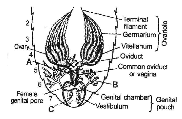
A-Spermatheca, B-Collaterial glands, C-Gonapophyses
A-Phallic gland, B-Collaterial glands, C-Gonapophyses
A-Spermatheca, B-Seminal vesicle, C-Gonapophyses
A-Spermatheca, B-Collateral glands, C-Tegmina
Lack of blood supply and presence of the noncellular basement membrane are the characteristics of the
muscular tissue
fluid connective tissue
epithelial tissue
nervous tissue