
Nervous tissue cells that play several supporting roles but do not transmit impulses are called
glial cells
dendrites
nerve cells
neurons
Correct Answer :
A. glial cells
The neuroglial cells are non-excitable cells that protect and support neurons.
Related Questions
The chondrocytes of connective tissue are
fibre secreting cells
bone forming cells
cartilage cells
bone eating cells
The given figure shows the nephridial system of earthworm and answer the question.
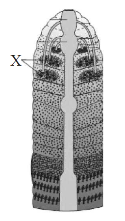
Select the option which shows the correct identification and the function of the structure marked as X.
Lateral heart. It is a blood pumping organ.
Calciferous glands. They neutralize the humic acid present in humus.
Nephridia. It regulates the volume and composition of the body fluids.
Blood glands. They produce blood cells and haemoglobin which is dissolved in blood plasma.
If the head of cockroach is cut off, it will still alive for as long as one week. It is because of
the body which is covered with a hard chitinous exoskeleton.
head which holds a bit of nervous system.
head which is of no use.
food capturing appratus which is found elsewhere.
The ciliated columnar epithelial cells in humans occur in
Eustachian tube and stomach lining
bronchioles and fallopian tubes
bile duct and oesophagus
fallopian tubes and urethra
In which one of the following preparations, cell junctions come across most frequently ?
Ligament
Tendon
Cartilage
Ciliated epithelium
Refer the given figure of female reproductive system of cockroach and identify the correct labels (marked as 1, 2, 3 and 4) which are collectively called genital pouch.
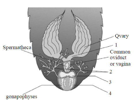
1 & 2
1 & 3
2 & 4
3 & 4
Match the types of connective tissue given in column-I with their examples given column-II and choose the correct option.
| Column-I | Column-II |
|---|---|
| (Types of connective) | (Examples) |
| A. Loose connective | I. Tendons and ligaments tissue |
| B. Dense regular | II. Skin tissue |
| C. Dense irregular | III. Cartilage, bones, blood tissue |
| D. Specialized | IV. Fibroblasts, macrophages connective tissue and mast cells |
A I; B IV; C II; D III
A I; B IV; C III; D II
A IV; B I; C II; D III
A IV; B II; C I; D III
Statement 1: Typhlosole increases the effective area of absorption in the intestine.
Statement 2: Typhlosole, present in the intestine, is the characteristic feature of cockroach.
Statement 1 and 2 are true and statement 2 is the correct explanation of statement 1.
Statement 1 and 2 are true and statement 2 is not the correct explanation of statement 1.
Both statements 1 and 2 are false.
Statement 1 is true and statement 2 is false.
Which of the following statement (s) is/are correct ?
- Loose connective tissue contains fibroblasts, macrophages and mast cells.
- Adipose tissue is a type of dense connective tissue located mainly beneath the skin.
- Tendons and ligaments are examples of dense irregular connective tissue.
- Cartilage, bones and blood are various types of specialized connective tissue.
Only (i)
Both (ii) and (iv)
Both (i) and (iii)
(i), (iii) and (iv)
Which of the following statement(s) regarding cell junctions is/are correct ?
Tight junctions help to stop substances from leaking across a tissue.
Adhering junctions perform cementing to keep neighbouring cells together.
Gap junctions facilitate the cells to communicate with each other by connecting the cytoplasm of adjoining cells, for rapid transfer of ions, small molecules and sometimes big molecules.
All of the above
Match the epithetial tissue given in column-I with its location given in column-II and choose the correct option.
| Column I | Column II |
|---|---|
| (Epithelial tissue) | (Location) |
| A. Cuboidal | I. Epidermis of skin |
| B. Ciliated | II. Inner lining of blood vessels |
| C. Columnar | III. Inner surface of gall bladder |
| D. Squamous | IV. Inner lining of fallopian tube |
| E. Keratinized | V. Lining of pancreatic duct squamous |
A V; B IV; C II; D III; E I
A III; B IV; C V; D II; E I
A V; B IV; C III; D II; E I
A III; B IV; C V; D I; E II
The following figures A, B and C are types of muscle tissue. Identify A, B and C.
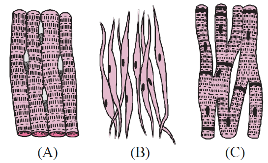
A Smooth muscle, B Cardiac muscle, C Skeletal muscle
A Skeletal muscle, B Smooth muscle, C Cardiac muscle
A Cardiac muscle, B Smooth muscle, C Skeletal muscle
A Smooth muscle, B Skeletal muscle , C Cardiac muscle
A student was given a sample of tissue. He observes and concludes the following characters.
- The cells are composed of a single layer of tall and slender cells.
- Their nuclei are located at the base.
- Free surface may have microvilli.
- It is found in the lining of stomach and intestine
- They help in secretion and absorption.
Based on the above features identify the epithelium.
Cuboidal epithelium
Columnar Epithelium
Squamous epithelium
Glandular epithelium
Which of the following type of muscle tissue is being described on the basis of given statements ?
- These muscle fibres taper at both ends and do not show striations.
- The wall of internal organs such as the blood vessels, stomach and intestine contain this type of muscle tissue.
- They are involuntary as their function cannot be directly controlled.
Skeletal muscle
Smooth muscle
Cardiac muscle
All of these
The intercellular material of the given figure is solid and resists compression. Identify the figure and the label marked as A & B.
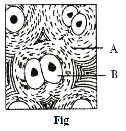
Fig - Cartilage, A - Collagen, B - Collagen
Fig - Cartilage, A - Microtubule, B - Collagen fibres
Fig - Bone, A - Chondrocyte, B - Chondroclast
Fig - Bone, A - Chondroclast, B - Osteoblast
Lack of blood supply and presence of the noncellular basement membrane are the characteristics of the
muscular tissue
fluid connective tissue
epithelial tissue
nervous tissue
The following figure shows the extarnal features of cockroach with few structures labelled as A, B, C, D, & E. Identify A to E .
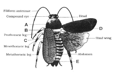
A- Mesothorax, B-Pronotum, C-Metathorax, D-Tegmina, E-Anal style
A- Pronotum, B-Metathorax, C-Mesothorax, D-Tegmina, E-Sterna
A- Pronotum, B-Mesothorax, C-Metathorax, D-Tegmina, E-Anal cerci
A- Pronotum, B-Mesothorax, C-Metathorax, D-Tegmina, E-Anal style
Cockroaches are brown or black bodied animals that are included in class _______ of phylum _______.
reptilia; annelida
insecta; arthropoda
insecta; annelida
reptilia; arthropoda
Read the following statements.
- It is a contractile tissue present only in the heart.
- Cell junctions fuse the plasma membranes of these cells and make them stick together.
- Communication juntions at some fusion points allow the cells to contract as a unit, i.e., when one cell receives a signal to contract, its neighbours are also stimulated to contract.
Which of the following type of tissue is being described by the above statements ?
Skeletal muscle
Cardiac muscle
Smooth muscle
Cartilage
Phallic organs in cockroach are related to
male excretory system.
male reproductive system.
female excretory system.
female reproductive system.
Which of the following animals maintain ecological balance?
Frog
Rabbit
Earthworm
Cockroach
Match the description given in column I with their examples given in column II and choose the correct option.
| Column I | Column II |
|---|---|
| (Description) | (Example) |
| 1. Aquatic respiratory | A. Skin organ |
| 2. Organ which acts | B. Ureter urogenital duct and opens into the cloaca |
| 3. A small median chamber | C. Cloaca that is used to pass faecal matter, urine and sperms to the exterior |
| 4. A triangular structure | D. Sinus venosus which joins the right atrium and receives blood through vena cava |
1 - A, 2 - B, 3 - C, 4 - D
1 - C, 2 - A, 3 - D, 4 - B
1 - B, 2 - A, 3 - C, 4 - D
1 - C, 2 - B, 3 - D, 4 - A
The major functions of loose connective tissue include
occupying spaces between organs and supporting epithelia.
supporting and surrounding blood vessels and nerves
cushioning organs, storing lipids and facilitating diffusion.
All of the above
The given figure shows open circulatory system of cockroach with structure marked as A, B and C. Which structure is a 13 pair of wing shaped involuntary muscles and mantain blood circulation?
A
B
C
Both A nad B
Which of the following statement(s) is/are correct regarding respiratory system of cockroach ?
- It consists of a network of trachea, that open through 12 pairs of small holes called spiracles present on the lateral side of the body.
- Thin branching tubes carry oxygen from the air to all the parts.
- The opening of the spiracles is regulated by sphincters.
- Exchange of gases take place at the tracheoles by diffusion.
Only (i)
(i), (ii) and (iii)
(ii), (iii) and (iv)
All of these
Match column-I (type of epithelium) with column-II (Description) and choose the correct option.
| Column-I | Column-II |
|---|---|
| (Types of epithelium) | (Description) |
| A. Squamous | I. It is composed of a epithelium single-layer of cube-like cells |
| B. Cuboidal | II. Having cilia on their free epithelium surface |
| C. Columnar | III. It is composed of a single epithelium layer of tall and slender cells |
| D. Ciliated | IV. It is made up of a single thin epithelium layer of flattened cells with irregular boundaries |
A IV; B I; C III; D II
A I; B IV; C III; D II
A IV; B I; C II; D III
A IV; B III; C I; D II
Read the following statements and answer the question.
- They have a hard and non-pliable ground substance rich in calcium salts and collagen fibres.
- They support and protect softer tissues and organs.
- Osteocytes are present in the spaces called lacunae.
- They also interact with skeletal muscles attached to them to bring about movements.
Which of the following type of tissue is being described by above statements ?
Cartilage
Bone
Blood
Neurons
Which of the following statement is correct regarding Female reproductive system of earthworm ?
It consists of two large ovaries, lying laterally in the 6th 7th abdominal segments.
Each ovary is formed of a group of five ovarian tubules or ovarioles, containing a chain of developing ova.
A pair of spermatheca is present in the 5th segment which opens into the genital chamber.
None of the above
The diagram given below represents the reproductive organ of male cockroach. Choose the correct labelling of the part of marked as A, B, C and D.
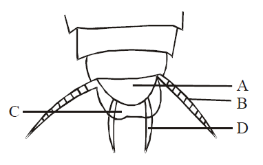
A 8th sternum, B - Anal cercus, C - 10th tergum, D - Anal style
A - 10th tergum, B - Anal cercus, C - Anal style, D - 8th sternum
A - Anal style, B - Anal cercus, C - 10th tergum, D- 8th sternum
A - Anal cercus, B - 8th sternum, C - 10th tergum, D - Anal style.
Which one of the following pairs of structures distinguishes a nerve cell from other types of cell ?
Vacuoles and Fibres
Flagellum and Medullary sheath
Nucleus and Mitochondria
Cell body and Dendrites