
The kind of epithelium which forms the inner walls of blood vessels is
cuboidal epithelium
columnar epithelium
ciliated columnar epithelium
squamous epithelium
Correct Answer :
D. squamous epithelium
Squamous epithelium is formed of thin discoidal and polygonal cells that fit like tiles in a floor, so is also called pavement epithelium. It is found in the walls of blood vessels, in the alveoli of lung for exchange of gases, and in Bowmans capsule of nephron for ultra filtration.
Related Questions
The intercellular material of the given figure is solid and resists compression. Identify the figure and the label marked as A & B.
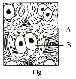
Fig - Cartilage, A - Collagen, B - Collagen
Fig - Cartilage, A - Microtubule, B - Collagen fibres
Fig - Bone, A - Chondrocyte, B - Chondroclast
Fig - Bone, A - Chondroclast, B - Osteoblast
Which of the follwing statement(s) is/are correct ?
- Cockroaches are brown or black bodied animals that are included in class insecta of phylum arthropoda.
- Males bear a pair of short, thread like anal styles which are absent in females.
- Heart of cockroach consists of elongated muscular tube lying along mid dorsal line of thorax and abdomen.
- The nymph grows by moulting about 13 times to reach the adult form.
Only (i)
Both (ii) and (iii)
Both (i) and (ii)
All of the above
Which of the following statement is incorrect regarding cuboidal epithelium ?
It is an epithelial tissue.
It is composed of a single layer of cube-like cells.
They are found in the walls of blood vessels and air sacs of lungs.
Secretion and absorption are the main functions of these tissue.
Which of the following is involved in the production of new blood cells ?
Adipose cell
Bone marrow
Liver
Matrix
The figure given below shows the head region of cockroach. Identify A to F.
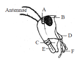
A- Compound eye, B-Ocellus, C-Maxilla, D-Mandible, E-Labrum, F-Labium
A- Ocellus, B-Compound eye, C-Mandible, D-Maxilla, E-Labrum, F-Labium
A- Ocellus, B-Compound eye, C-Mandible, D-Maxilla, E-Labium, F-Labrum
A- Ocellus, B-Compound eye, C-Maxilla, D-Mandible, E-Labrum, F-Labium
Which of the following type of tissue is being described by the given statements ?
- They are named because of their special function of linking and supporting other tissues/organs of the body.
- They include cartilage, bone, adipose and blood.
- They provide strength, elasticity and flexibility to the tissue.
- They also secrete modified polysaccharides, which accumulate between cells and fibres and act as matrix.
Epithelial tissue
Connective tissue
Muscle tissue
Neural tissue
Statement 1 : Cartilage (protein matrix) and bone (calcium matrix) are rigid connective tissue.
Statement 2 : Blood is connective tissue in which plasma is the matrix.
Statement- 1 and statement-2 are true and statement-2 is a correct explanation for statement -1
Statement -1 and statement-2 are true and statement-2 is not a correct explanation for statement -1
Statement - 1 is true and statement- 2 is false
Both the statements are false.
Smooth muscles are______.
voluntary, branched, uninucleate
voluntary, multinucleate, cylindrical
involuntary, cylindrical, multinucleate
involuntary, spindle shaped, uninucleated, tapering
Refer the given figure of female reproductive system of cockroach and identify the correct labels (marked as 1, 2, 3 and 4) which are collectively called genital pouch.
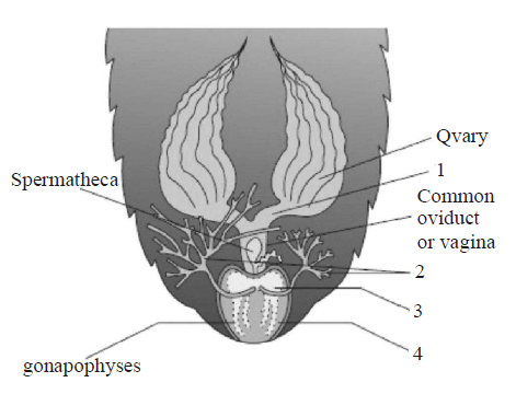
1 & 2
1 & 3
2 & 4
3 & 4
Figure given below shows reproductive system of female cockroach. The correct labellings indicated by alphabets (A, B & C) are respectively
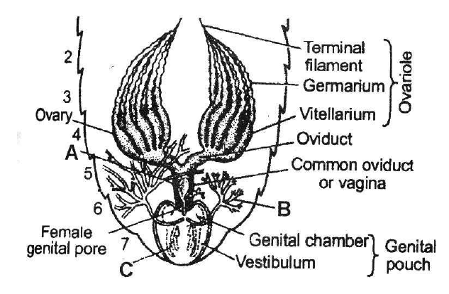
A-Spermatheca, B-Collaterial glands, C-Gonapophyses
A-Phallic gland, B-Collaterial glands, C-Gonapophyses
A-Spermatheca, B-Seminal vesicle, C-Gonapophyses
A-Spermatheca, B-Collateral glands, C-Tegmina
Which of the following statement(s) is/are correct regarding respiratory system of cockroach ?
- It consists of a network of trachea, that open through 12 pairs of small holes called spiracles present on the lateral side of the body.
- Thin branching tubes carry oxygen from the air to all the parts.
- The opening of the spiracles is regulated by sphincters.
- Exchange of gases take place at the tracheoles by diffusion.
Only (i)
(i), (ii) and (iii)
(ii), (iii) and (iv)
All of these
Match column-I (type of epithelium) with column-II (Description) and choose the correct option.
| Column-I | Column-II |
|---|---|
| (Types of epithelium) | (Description) |
| A. Squamous | I. It is composed of a epithelium single-layer of cube-like cells |
| B. Cuboidal | II. Having cilia on their free epithelium surface |
| C. Columnar | III. It is composed of a single epithelium layer of tall and slender cells |
| D. Ciliated | IV. It is made up of a single thin epithelium layer of flattened cells with irregular boundaries |
A IV; B I; C III; D II
A I; B IV; C III; D II
A IV; B I; C II; D III
A IV; B III; C I; D II
Which of the following statement (s) is/are correct ?
- Loose connective tissue contains fibroblasts, macrophages and mast cells.
- Adipose tissue is a type of dense connective tissue located mainly beneath the skin.
- Tendons and ligaments are examples of dense irregular connective tissue.
- Cartilage, bones and blood are various types of specialized connective tissue.
Only (i)
Both (ii) and (iv)
Both (i) and (iii)
(i), (iii) and (iv)
Which of the following statement(s) regarding cell junctions is/are correct ?
Tight junctions help to stop substances from leaking across a tissue.
Adhering junctions perform cementing to keep neighbouring cells together.
Gap junctions facilitate the cells to communicate with each other by connecting the cytoplasm of adjoining cells, for rapid transfer of ions, small molecules and sometimes big molecules.
All of the above
Which of the following type of cell junction is not found in animal tissues ?
Adhering junction
Tight junction
Gap junction
Plasmodesmata
Which of the following types of connective tissue is mismatched with its matrix ?
Areolar Loosely packed matrix of protein fibres
Bone Mineralized matrix
Cartilage Highly vascular matrix
Blood Liquid matrix
Non-ciliated simple columnar epithelium often contains _______, which increase the surface area for secretion and absorption.
flagella
collagen fibres
microvilli
all of these
Neuroglia are
excitable cells of neural tissue.
supporting and non-excitable cells of neural tissue.
two to three times in volume of neural tissue.
protective and excitable cells of neural tissue.
Lack of blood supply and presence of the noncellular basement membrane are the characteristics of the
muscular tissue
fluid connective tissue
epithelial tissue
nervous tissue
Which of the following animals maintain ecological balance?
Frog
Rabbit
Earthworm
Cockroach
Which of the following statement(s) is/are correct regarding compound epithelium ?
It is made of more than one layer of cells and thus has a limited role in secretion and absorption.
Their main function is to provide protection against chemical and mechanical stresses.
They cover the dry surface of the skin, moist surface of buccal cavity, pharynx, inner lining of ducts of salivary glands and pancreatic ducts.
All of the above
Match the epithetial tissue given in column-I with its location given in column-II and choose the correct option.
| Column I | Column II |
|---|---|
| (Epithelial tissue) | (Location) |
| A. Cuboidal | I. Epidermis of skin |
| B. Ciliated | II. Inner lining of blood vessels |
| C. Columnar | III. Inner surface of gall bladder |
| D. Squamous | IV. Inner lining of fallopian tube |
| E. Keratinized | V. Lining of pancreatic duct squamous |
A V; B IV; C II; D III; E I
A III; B IV; C V; D II; E I
A V; B IV; C III; D II; E I
A III; B IV; C V; D I; E II
The following figure shows the extarnal features of cockroach with few structures labelled as A, B, C, D, & E. Identify A to E .
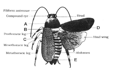
A- Mesothorax, B-Pronotum, C-Metathorax, D-Tegmina, E-Anal style
A- Pronotum, B-Metathorax, C-Mesothorax, D-Tegmina, E-Sterna
A- Pronotum, B-Mesothorax, C-Metathorax, D-Tegmina, E-Anal cerci
A- Pronotum, B-Mesothorax, C-Metathorax, D-Tegmina, E-Anal style
The given figure shows open circulatory system of cockroach with structure marked as A, B and C. Which structure is a 13 pair of wing shaped involuntary muscles and mantain blood circulation?
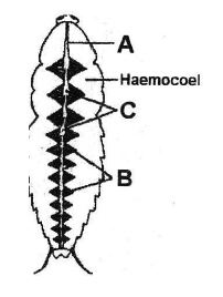
A
B
C
Both A nad B
In the given diagram of areolar connective tissue, the different cells and parts have been marked by alphabets (A, B, C & D). Choose the answer in which these alphabets correctly match with the parts and cells they indicate.
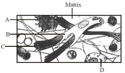
A-Adipocyte, B-Collagen fibres, C-Microfilament, D-Mast cells
A-Macrophage, B-Collagen fibres, C-Microfilament, D-Mast cells
A-Macrophage, B-Collagen fibres, C-Microtubule, D-RBC
A-Macrophage, B-Fibroblast, C-Collagen fibres, D-Mast cells
Match column-I with column-II and choose the correct option.
| Column-I | Column-II |
|---|---|
| A. Periplaneta | I. Hepatic caecae americana |
| B. A ring of 6-8 blind | II. Phylum arthropoda tubules |
| C. Vascular system | III. Spiracles |
| D. 10 pairs of small | IV. Malpighian tubules holes |
| E. Excretion | V. Open type |
A I; B II; C III; D IV; E V
A II; B I; C V; D III; E IV
A II; B I; C III; D V; E IV
A III; B IV; C II; D V; E I
A student was given a sample of two tissues. He observes the tissues under the microscope and draws their figures (1 and 2) as shown below.
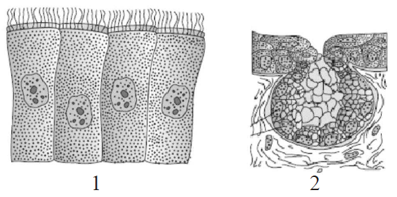
Identify the tissues (1 and 2).
1: Columnar cells bearing cilia; 2: Unicellular glandular epithelium
1: Cuboidal cells bearing cilia; 2: Multicellular glandular epithelium
1: Compound cells bearing cilia; 2: Unicellular glandular epithelium
1: Columnar cells bearing cilia; 2: Multicellular glandular epithelium
Read the following statements.
- It is a contractile tissue present only in the heart.
- Cell junctions fuse the plasma membranes of these cells and make them stick together.
- Communication juntions at some fusion points allow the cells to contract as a unit, i.e., when one cell receives a signal to contract, its neighbours are also stimulated to contract.
Which of the following type of tissue is being described by the above statements ?
Skeletal muscle
Cardiac muscle
Smooth muscle
Cartilage
Three essential components of most neurons are
simple epithelium, extracellular matrix and nerves.
axon, dendrites and cell body.
nerve cells, synapse and neuroglia.
mylein sheeth, node of Ranvier and Schwann cells.
A student was given a sample of tissue. He observes and concludes the following characters.
- The cells are composed of a single layer of tall and slender cells.
- Their nuclei are located at the base.
- Free surface may have microvilli.
- It is found in the lining of stomach and intestine
- They help in secretion and absorption.
Based on the above features identify the epithelium.
Cuboidal epithelium
Columnar Epithelium
Squamous epithelium
Glandular epithelium